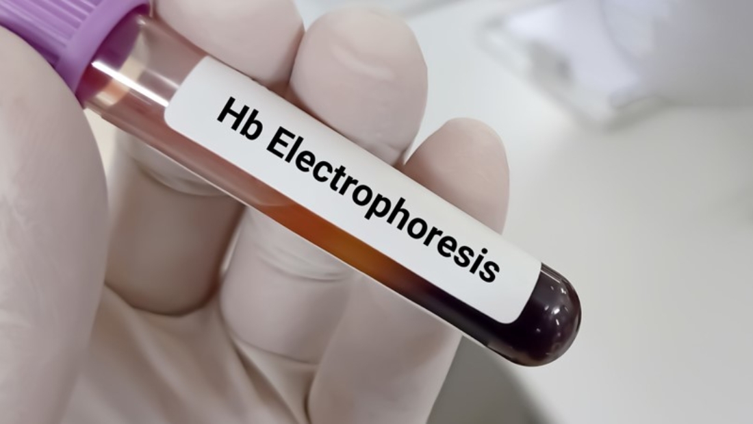
60-1024x536

How Blood Tests Detect Infectious Diseases: From HIV to Hepatitis.

Introduction

Infectious diseases have shaped human history, from the bubonic plague to modern-day pandemics. The invisible enemies—viruses, bacteria, fungi, and parasites—can spread quickly and silently. One of the most powerful tools we have in detecting, managing, and controlling these threats is the humble blood test.

Blood is more than just a red fluid coursing through our veins. It is a dynamic, information-rich medium that reflects the body’s internal state. Through blood testing, clinicians can unearth the fingerprints of pathogens, determine the stage of an infection, monitor response to treatment, and even detect asymptomatic carriers. This is particularly crucial for blood-borne infections like HIV (Human Immunodeficiency Virus) and hepatitis viruses (A, B, C, D, and E).

In this blog, we’ll take a deep dive into the science behind how blood tests detect infectious diseases—focusing especially on HIV and the hepatitis virus family—while also touching upon emerging technologies, challenges, and public health implications.


Chapter 1: The Science of Blood Testing

What Is a Blood Test?

At its core, a blood test involves collecting a sample of blood—usually from a vein in the arm—and analyzing it in a laboratory for specific biomarkers. These biomarkers could be anything from white blood cell counts, antibodies, enzymes, or even fragments of a pathogen’s genetic material.

Blood tests fall into two main categories:

  • Serological tests – detect antibodies or antigens.

  • Molecular tests – detect the genetic material (RNA or DNA) of the pathogen.

Each type of test offers different advantages in terms of accuracy, timing, and application.


Chapter 2: Types of Blood Tests for Infectious Diseases

1. Antibody Tests (Serology)

These tests look for antibodies that the immune system produces in response to an infection. They can confirm whether a person has been exposed to a pathogen in the past or is currently infected.

  • IgM antibodies suggest recent infection.

  • IgG antibodies indicate past exposure or long-standing infection.

2. Antigen Tests

Antigens are parts of the pathogen itself (often proteins). Detecting antigens in the blood is useful for identifying active infections.

3. PCR (Polymerase Chain Reaction) and NAAT (Nucleic Acid Amplification Tests)

These are molecular tests that identify genetic material from viruses or bacteria. They’re extremely sensitive and often used for early detection.

4. Viral Load Tests

These tests measure the amount of virus in the blood. They are critical for monitoring diseases like HIV and hepatitis C.

5. CD4 Count and Liver Enzymes

Used in HIV and hepatitis respectively, these are not direct pathogen tests but help assess immune function or organ damage caused by the infection.


Chapter 3: Detecting HIV Through Blood Tests

How HIV Works

HIV targets and destroys CD4 T cells, crucial components of the immune system. As the virus replicates, the immune system weakens, leaving the body vulnerable to opportunistic infections.

Key Blood Tests for HIV

1. HIV Antibody Test

  • Window period: 3 to 12 weeks post-exposure.

  • Common forms include ELISA (enzyme-linked immunosorbent assay).

2. HIV Antigen/Antibody Combination Test (4th generation test)

  • Detects both p24 antigen and HIV antibodies.

  • Shorter window period (2 to 4 weeks).

  • Highly sensitive and standard in most HIV screening protocols.

3. HIV RNA Test

  • Direct detection of HIV’s genetic material.

  • Can detect infection as early as 10 days after exposure.

  • Used for early diagnosis and in neonates born to HIV-positive mothers.

4. CD4 Count

  • Reflects immune system health.

  • Normal range: 500–1,600 cells/mm³. Treatment typically begins below 500.

5. HIV Viral Load Test

  • Quantifies virus in the blood.

  • Important for monitoring response to antiretroviral therapy (ART).

HIV Testing Strategy

  • Initial screening: Antigen/antibody test.

  • Confirmation: HIV-1/HIV-2 differentiation assay or RNA test.

  • Follow-up: CD4 and viral load monitoring.


Chapter 4: Detecting Hepatitis Through Blood Tests

The Hepatitis Virus Family

  • Hepatitis A: Fecal-oral route. Acute infection only.

  • Hepatitis B: Bloodborne/sexual transmission. Can be acute or chronic.

  • Hepatitis C: Bloodborne. Often chronic.

  • Hepatitis D: Requires co-infection with hepatitis B.

  • Hepatitis E: Fecal-oral route. Mostly acute.

Key Blood Tests by Hepatitis Type

Hepatitis A (HAV)

  • Anti-HAV IgM: Indicates acute infection.

  • Anti-HAV IgG: Indicates past infection or immunity.

Hepatitis B (HBV)

  • HBsAg (Hepatitis B surface antigen): Indicates current infection.

  • Anti-HBs: Sign of immunity (via vaccine or past infection).

  • Anti-HBc IgM: Recent infection.

  • Anti-HBc IgG: Past or chronic infection.

  • HBV DNA test: Measures viral load.

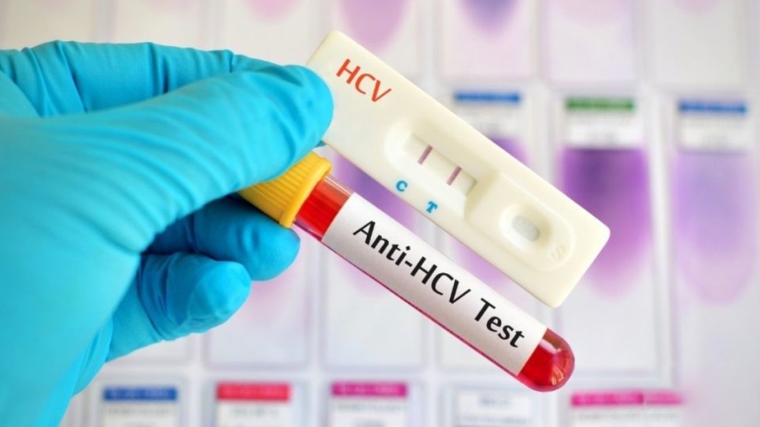
Hepatitis C (HCV)

  • Anti-HCV antibodies: Indicates exposure.

  • HCV RNA PCR test: Confirms active infection.

  • HCV Genotyping: Important for guiding treatment.

Hepatitis D (HDV)

  • Detected via Anti-HDV antibodies and HDV RNA.

  • Only occurs in the presence of HBV.

Hepatitis E (HEV)

  • Anti-HEV IgM: Acute infection.

  • Anti-HEV IgG: Previous exposure or immunity.

  • HEV RNA: Confirms infection, especially in immunocompromised individuals.


Chapter 5: Window Periods and Why They Matter

The window period is the time between initial infection and when a test can reliably detect it. This is critical because:

  • False negatives may occur during this time.

  • Transmission risk is high despite negative results.

Examples:

  • HIV antibody test: 3–12 weeks.

  • 4th gen HIV test: 2–4 weeks.

  • HBsAg: Appears 1–10 weeks after exposure.

  • Anti-HCV: 8–11 weeks; HCV RNA can be detected earlier.


Chapter 6: Blood Screening in Public Health

Blood Donation Safety

All donated blood is screened for:

  • HIV

  • Hepatitis B and C

  • Syphilis

  • HTLV (Human T-lymphotropic virus)

  • West Nile virus (in certain regions)

Infection Surveillance

Blood testing helps health authorities:

  • Monitor outbreaks.

  • Identify transmission patterns.

  • Implement prevention campaigns.


Chapter 7: Advances in Blood Testing Technology

1. Rapid Diagnostic Tests (RDTs)

  • Provide results in under 30 minutes.

  • Useful in remote or resource-limited settings.

2. Multiplex Assays

  • Test for multiple pathogens simultaneously.

  • Save time and resources.

3. Point-of-Care Testing

  • Performed at the patient’s location.

  • Useful for quick decision-making.

4. Next-Generation Sequencing (NGS)

  • Can detect unknown or multiple pathogens.

  • Valuable in research and outbreak investigations.

5. CRISPR-based Diagnostics

  • Ultra-sensitive.

  • Future potential for real-time, low-cost testing.


Chapter 8: Challenges and Limitations

False Negatives and False Positives

No test is perfect. Issues may arise due to:

  • Window periods.

  • Technical errors.

  • Cross-reactivity.

Access to Testing

  • Many populations lack access due to cost, stigma, or geography.

Need for Follow-up

Positive results often require:

  • Confirmatory testing.

  • Further evaluation (e.g., liver biopsy, genotyping).


Chapter 9: Ethical and Social Implications

  • Informed consent is essential before testing.

  • Stigma can deter individuals from seeking testing.

  • Confidentiality must be strictly maintained.

  • Counseling should accompany diagnosis, especially for lifelong infections like HIV.


Chapter 10: Future Directions

Personalized Medicine

Tailoring treatment based on viral load, genotype, and immune markers.

AI and Automation

Machine learning may soon enhance diagnostic accuracy and pattern recognition in blood test data.

Global Health Integration

Combining blood testing programs with vaccination, education, and harm reduction can drastically curb the spread of infectious diseases.


Conclusion

Blood tests have revolutionized our ability to detect and combat infectious diseases. From the early detection of HIV to the precise management of hepatitis, these diagnostic tools serve as the cornerstone of modern medicine. But to maximize their potential, we must ensure they are accessible, accurate, and ethically administered.

Whether you’re a healthcare professional, policy maker, or simply a concerned citizen, understanding how blood tests work empowers you to make informed decisions—for yourself and your community.

Laboratory Equipment

The Latest Innovations in Laboratory Equipment for 2025

The Latest Innovations in Laboratory Equipment for 2025.

Introduction

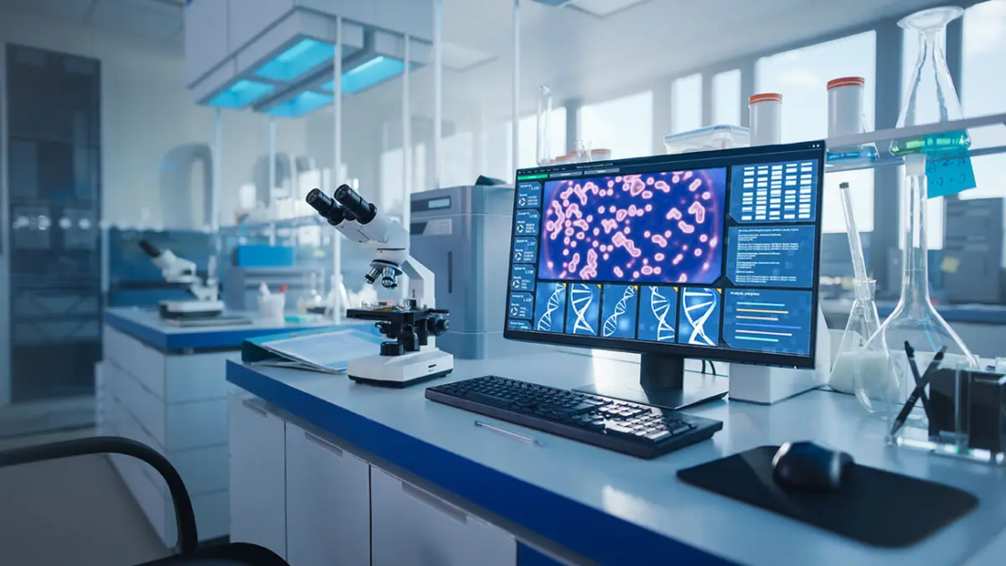
The world of science and research is constantly evolving, and with it, the tools and equipment that researchers rely on to make groundbreaking discoveries. As we look ahead to 2025, the landscape of laboratory equipment is set to undergo some exciting transformations. From advanced automation to cutting-edge analytical instruments, the latest innovations are poised to revolutionize the way we conduct experiments, analyze data, and push the boundaries of human knowledge.

In this blog, we’ll take a deep dive into the most promising advancements in laboratory equipment for 2025. Whether you’re a seasoned researcher, a lab manager, or simply someone with a passion for science, this comprehensive guide will give you a glimpse into the future of laboratory technology. So, let’s roll up our sleeves and explore the innovations that are set to shape the future of research.


1. Automation and Robotics: The Rise of Smart Labs

1.1 Fully Automated Workstations

By 2025, laboratories are expected to become increasingly automated, with fully automated workstations taking center stage. These systems are designed to handle repetitive tasks such as pipetting, sample preparation, and data entry, freeing up researchers to focus on more complex and creative aspects of their work. Companies like Thermo Fisher Scientific and Hamilton Robotics are leading the charge, developing robotic systems that can perform a wide range of tasks with precision and efficiency.

1.2 AI-Powered Lab Assistants

Artificial intelligence (AI) is making its way into the lab, with AI-powered lab assistants becoming a reality. These systems can analyze data, predict outcomes, and even suggest experimental protocols based on historical data. For example, AI algorithms can optimize reaction conditions in chemical synthesis or identify potential drug candidates in pharmaceutical research. By 2025, we can expect AI to become an integral part of laboratory workflows, enhancing productivity and accuracy.

1.3 Collaborative Robots (Cobots)

Collaborative robots, or cobots, are designed to work alongside human researchers, providing assistance with tasks that require precision and repeatability. Unlike traditional industrial robots, cobots are equipped with advanced sensors and safety features, making them safe to operate in close proximity to humans. In 2025, cobots are expected to become more affordable and accessible, enabling even small labs to benefit from robotic automation.


2. Next-Generation Analytical Instruments

2.1 High-Resolution Mass Spectrometers

Mass spectrometry is a cornerstone of analytical chemistry, and by 2025, we can expect to see even more advanced high-resolution mass spectrometers. These instruments will offer unparalleled sensitivity and accuracy, enabling researchers to detect and analyze trace amounts of compounds with ease. Innovations in ion mobility spectrometry and tandem mass spectrometry will further enhance the capabilities of these instruments, opening up new possibilities in proteomics, metabolomics, and environmental analysis.

2.2 Portable Spectrometers

The demand for portable analytical instruments is on the rise, and by 2025, portable spectrometers will become more compact, affordable, and user-friendly. These devices will allow researchers to perform on-site analysis in remote locations, from monitoring environmental pollutants to diagnosing diseases in the field. Companies like Agilent Technologies and Bruker are already developing handheld spectrometers that deliver lab-quality results in a portable format.

2.3 Quantum Sensors

Quantum technology is set to revolutionize analytical instrumentation, with quantum sensors offering unprecedented levels of precision. These sensors leverage the principles of quantum mechanics to detect minute changes in magnetic fields, temperature, and pressure. By 2025, quantum sensors are expected to find applications in a wide range of fields, from medical diagnostics to materials science.


3. Advanced Imaging Technologies

3.1 Super-Resolution Microscopy

Super-resolution microscopy has already transformed the field of cell biology, and by 2025, we can expect even more advanced imaging techniques. These technologies will enable researchers to visualize cellular structures and processes at the nanoscale, providing new insights into the mechanisms of life. Innovations in fluorescence microscopy and 3D imaging will further enhance the capabilities of these instruments.

3.2 Cryo-Electron Microscopy (Cryo-EM)

Cryo-EM has emerged as a powerful tool for studying the structure of proteins and other biomolecules. By 2025, advancements in cryo-EM technology will make it faster, more accessible, and more affordable. Improved detectors and software algorithms will enable researchers to obtain high-resolution images in a fraction of the time, accelerating discoveries in structural biology.

3.3 Multimodal Imaging Systems

Multimodal imaging systems combine multiple imaging techniques, such as MRI, CT, and PET, into a single platform. By 2025, these systems will become more integrated and user-friendly, allowing researchers to obtain comprehensive data from a single experiment. This will be particularly valuable in medical research, where multimodal imaging can provide a more complete picture of complex diseases.


4. Lab-on-a-Chip and Microfluidics

4.1 Miniaturized Diagnostic Devices

Lab-on-a-chip technology is revolutionizing diagnostics, enabling rapid and cost-effective analysis of biological samples. By 2025, we can expect to see even more advanced miniaturized diagnostic devices that can detect diseases, monitor biomarkers, and perform genetic testing with just a drop of blood. These devices will be particularly valuable in resource-limited settings, where access to traditional laboratory equipment is limited.

4.2 Organ-on-a-Chip Systems

Organ-on-a-chip systems are designed to mimic the structure and function of human organs, providing a powerful tool for drug development and toxicity testing. By 2025, these systems will become more sophisticated, with the ability to replicate complex organ interactions and disease states. This will enable researchers to conduct more accurate and ethical preclinical studies, reducing the need for animal testing.

4.3 Microfluidic Drug Delivery Systems

Microfluidics is also making waves in the field of drug delivery, with the development of targeted and controlled-release systems. By 2025, we can expect to see more advanced microfluidic devices that can deliver drugs directly to specific tissues or cells, minimizing side effects and improving therapeutic outcomes.


5. Sustainable and Eco-Friendly Lab Equipment

5.1 Energy-Efficient Instruments

As the world becomes more environmentally conscious, laboratories are also making efforts to reduce their carbon footprint. By 2025, we can expect to see more energy-efficient lab equipment, from refrigerators and freezers to analytical instruments. These devices will be designed to consume less power without compromising performance, helping labs reduce their energy costs and environmental impact.

5.2 Biodegradable Consumables

The use of single-use plastics in laboratories has come under scrutiny, and by 2025, we can expect to see a shift towards biodegradable and recyclable consumables. Companies are already developing eco-friendly alternatives to traditional lab plastics, such as pipette tips, petri dishes, and centrifuge tubes made from plant-based materials.

5.3 Waterless Labs

Water is a precious resource, and laboratories are among the largest consumers of water. By 2025, we can expect to see the emergence of waterless labs, where traditional water-intensive processes are replaced with dry or low-water alternatives. For example, waterless autoclaves and air-cooled condensers are already being developed to reduce water usage in labs.


6. Enhanced Data Management and Connectivity

6.1 Cloud-Based Lab Management Systems

The future of lab management lies in the cloud, with cloud-based systems enabling seamless data sharing and collaboration. By 2025, we can expect to see more labs adopting cloud-based platforms for managing experiments, tracking samples, and analyzing data. These systems will offer enhanced security, scalability, and accessibility, making it easier for researchers to work together across different locations.

6.2 Internet of Things (IoT) in the Lab

The Internet of Things (IoT) is making its way into the lab, with connected devices enabling real-time monitoring and control of experiments. By 2025, we can expect to see more IoT-enabled lab equipment, from smart incubators to connected spectrophotometers. These devices will provide researchers with valuable insights into their experiments, helping them optimize conditions and improve reproducibility.

6.3 Blockchain for Data Integrity

Blockchain technology is set to play a key role in ensuring data integrity and transparency in research. By 2025, we can expect to see more labs using blockchain to securely store and share data, ensuring that results are tamper-proof and verifiable. This will be particularly valuable in fields like clinical research, where data integrity is critical.


7. Personalized and Precision Medicine Tools

7.1 Next-Generation Sequencing (NGS)

Next-generation sequencing (NGS) has revolutionized genomics, and by 2025, we can expect even more advanced NGS platforms. These systems will offer faster sequencing speeds, higher accuracy, and lower costs, enabling researchers to sequence entire genomes in a matter of hours. This will drive advancements in personalized medicine, where treatments are tailored to an individual’s genetic makeup.

7.2 CRISPR-Based Diagnostics

CRISPR technology is not just for gene editing—it’s also being used for diagnostics. By 2025, we can expect to see more CRISPR-based diagnostic tools that can detect diseases with high specificity and sensitivity. These tools will be particularly valuable for detecting infectious diseases and genetic disorders.

7.3 Wearable Health Monitors

Wearable health monitors are becoming increasingly sophisticated, and by 2025, they will play a key role in personalized medicine. These devices will enable continuous monitoring of vital signs, biomarkers, and other health metrics, providing researchers with valuable data for studying disease progression and treatment outcomes.


Conclusion: The Future of Laboratory Equipment

As we look ahead to 2025, it’s clear that the world of laboratory equipment is on the brink of a revolution. From automation and AI to advanced imaging and sustainable practices, the latest innovations are set to transform the way we conduct research and make discoveries. These advancements will not only enhance the efficiency and accuracy of laboratory workflows but also open up new possibilities for scientific exploration.

Whether you’re a researcher, a lab manager, or simply someone with a passion for science, the future of laboratory equipment holds something for everyone. By embracing these innovations, we can push the boundaries of what’s possible and continue to make meaningful contributions to the world of science and beyond.

So, here’s to the future—a future where laboratories are smarter, more efficient, and more sustainable than ever before. The possibilities are endless, and the journey is just beginning.

wbc

What is WBC and Its Function?

What is WBC and Its Function?

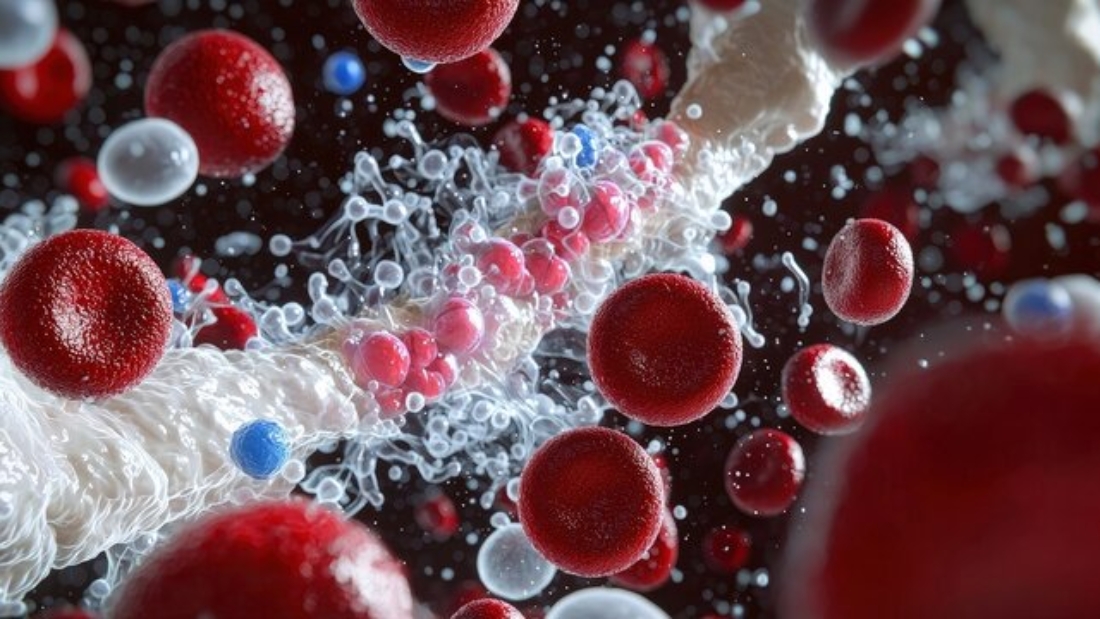
When we think about the incredible complexity of the human body, one of the most remarkable components is its ability to protect itself from harm. A key player in this protective mechanism is the White Blood Cell (WBC), also known as leukocyte. Understanding WBCs and their functions is essential to appreciating how the body combats infections and maintains overall health.

What are WBCs?

White blood cells are an integral part of the immune system. Unlike red blood cells, which primarily transport oxygen, WBCs are involved in defending the body against infections, foreign invaders, and abnormal cells. These cells are produced in the bone marrow and are found in blood and lymphatic tissues. While they make up only about 1% of the blood, their impact on health is immense.

WBCs are not a single type of cell but rather a group of diverse cells, each with specialized roles in immune defense. The primary types of WBCs include:

  1. Neutrophils
  2. Lymphocytes (B cells, T cells, and Natural Killer cells)
  3. Monocytes
  4. Eosinophils
  5. Basophils

Each type plays a unique role in maintaining immunity and combating disease.

Functions of WBCs

The primary function of WBCs is to protect the body from infections, but their specific roles go far beyond that. Here’s a closer look at how each type of WBC contributes to overall health, with examples:

  1. Neutrophils:
    • Neutrophils are the most abundant type of WBC and the first responders to microbial infections.
    • They are especially effective against bacterial and fungal infections.
    • Example: If you get a cut that becomes infected, neutrophils are the first to arrive at the wound site to engulf and destroy bacteria through a process called phagocytosis.
    • Additional Note: Neutrophils have a short lifespan (around 6 hours to a few days) and are continuously produced to ensure a ready defense.
  2. Lymphocytes:
    • Lymphocytes are key players in the adaptive immune response.
    • B cells: Produce antibodies that neutralize pathogens or mark them for destruction.
      • Example: After receiving a vaccine, B cells create antibodies to recognize and fight the specific virus in the future.
    • T cells: Help directly kill infected or cancerous cells and regulate immune responses.
      • Example: T cells attack and destroy cells infected with the flu virus.
    • Natural Killer (NK) cells: Provide rapid responses to viral infections and detect and control early signs of cancer.
      • Example: NK cells can identify and destroy cells infected by the herpes virus before symptoms appear.
    • Additional Note: Lymphocytes are long-lived cells, with some persisting for years as memory cells, ready to respond quickly to previously encountered pathogens.
  3. Monocytes:
    • Monocytes transform into macrophages and dendritic cells when they leave the bloodstream.
    • Example: Macrophages in the lungs engulf and break down dust particles or bacteria inhaled through the air.
    • Dendritic cells present antigens to T cells, initiating an immune response.
    • Additional Note: Monocytes also play a role in tissue repair by clearing dead cells and releasing growth factors.
  4. Eosinophils:
    • Eosinophils are crucial in combating parasitic infections and are involved in allergic reactions.
    • Example: If you have a parasitic worm infection, eosinophils release toxic proteins to kill the parasite and signal other immune cells to the site.
    • Additional Note: In allergic conditions, eosinophils can contribute to inflammation, as seen in asthma and hay fever.
  5. Basophils:
    • Basophils play a role in allergic and inflammatory responses.
    • Example: During an allergic reaction to pollen, basophils release histamine, which causes symptoms like a runny nose and watery eyes.
    • Additional Note: Basophils work closely with mast cells to amplify the body’s inflammatory response, which can be protective but may also lead to discomfort in allergic conditions.

Importance of WBCs in Health

A healthy WBC count is critical for maintaining immunity. The normal range for WBCs in adults is typically between 4,000 and 11,000 cells per microliter of blood. Abnormal WBC counts can indicate health issues:

  • Leukocytosis (high WBC count): May indicate infections, inflammation, stress, or diseases like leukemia.
    • Example: A person with pneumonia may have elevated WBC levels as the body fights off the lung infection.
    • Additional Note: Temporary leukocytosis can also occur during intense physical activity or stress as the body ramps up defense mechanisms.
  • Leukopenia (low WBC count): Can result from conditions such as autoimmune diseases, bone marrow disorders, or severe infections.
    • Example: A patient undergoing chemotherapy may experience leukopenia, making them more susceptible to infections.
    • Additional Note: Persistent leukopenia requires close medical monitoring to prevent life-threatening infections.

Regular check-ups and blood tests help monitor WBC levels and detect underlying health issues early.

How to Support WBC Function

Maintaining a robust immune system involves supporting your WBCs through lifestyle and dietary choices:

  1. Nutrition: Consume a balanced diet rich in vitamins (like C, D, and E), minerals (like zinc and selenium), and antioxidants.
    • Example: Eating citrus fruits, spinach, and almonds provides essential nutrients for WBC health.
    • Additional Tip: Foods like garlic and turmeric have natural immune-boosting properties.
  2. Exercise: Regular physical activity improves circulation, helping WBCs reach areas of need more effectively.
    • Example: Moderate exercise, like brisk walking, enhances immune cell activity without overexertion.
    • Additional Tip: Avoid overtraining, as it can temporarily suppress WBC function.
  3. Sleep: Adequate sleep is crucial for immune regulation.
    • Example: Chronic sleep deprivation reduces WBC efficiency, increasing susceptibility to colds.
    • Additional Tip: Aim for 7-9 hours of quality sleep each night to optimize immune health.
  4. Stress Management: Chronic stress can suppress WBC production and function.
    • Example: Practices like yoga or meditation help reduce cortisol levels, which can impair WBC activity.
    • Additional Tip: Engaging in hobbies and spending time with loved ones can also lower stress.
  5. Avoid Smoking and Limit Alcohol: These habits can impair immune cell activity.
    • Example: Quitting smoking improves overall immune health and WBC function.
    • Additional Tip: Replace these habits with healthier alternatives, like drinking herbal teas or engaging in physical activities.

Conclusion

White blood cells are an essential component of the body’s immune system, tirelessly working to protect against infections and maintain internal balance. Understanding the roles and importance of WBCs not only highlights the sophistication of the human body but also underscores the value of a healthy lifestyle in supporting immune function. By prioritizing nutrition, exercise, and self-care, you can help your WBCs function optimally, ensuring better protection and overall well-being.

Cell Counter

What is a Cell Counter?

  1. Introduction

    • Definition of cell counters
    • Importance in medical and research laboratories
    • Brief history of cell counting methods
  2. Types of Cell Counters

    • Manual cell counters
    • Automated cell counters
    • Flow cytometers
    • Image-based cell counters
  3. How Cell Counters Work

    • Principles of operation
    • Key components
    • Step-by-step process of cell counting
  4. Applications of Cell Counters

    • Clinical diagnostics
    • Research applications
    • Pharmaceutical industry
    • Quality control in biotechnology
  5. Choosing the Right Cell Counter

    • Factors to consider
    • Comparison of different types
    • Cost vs. benefits
  6. Maintenance and Calibration

    • Importance of regular maintenance
    • Step-by-step calibration guide
    • Troubleshooting common issues
  7. Future Trends in Cell Counting Technology

    • Advancements in automation
    • Integration with AI and machine learning
    • The future of cell counting in personalized medicine
  8. Conclusion

    • Summary of key points
    • Final thoughts on the importance of cell counters

Blog Post: About Cell Counters: Understanding Their Importance in Modern Laboratories

Introduction

In the rapidly evolving world of medical and scientific research, cell counters play a pivotal role in ensuring accurate and efficient analysis of cell samples. A cell counter is a specialized instrument designed to quantify the number of cells in a given sample, whether it’s blood, tissue, or cultured cells. The significance of cell counters cannot be overstated; they are essential tools in various fields, including clinical diagnostics, research, and biotechnology.

Cell counting has a rich history, evolving from manual techniques that relied on microscope counting chambers to sophisticated automated systems capable of analyzing thousands of cells in mere seconds. This evolution has greatly enhanced the accuracy and reliability of cell counts, allowing researchers and healthcare professionals to make informed decisions based on precise data.

As we delve deeper into the world of cell counters, we will explore the different types available, how they work, their applications, and future trends in this crucial technology.


1. Types of Cell Counters

Cell counters can be categorized into several types, each designed to meet specific needs within laboratories:

  • Manual Cell Counters: Traditional methods using a hemocytometer allow for visual counting of cells under a microscope. While accurate, these methods are time-consuming and prone to human error.
  • Automated Cell Counters: These devices streamline the counting process by using advanced technology to provide fast and accurate results. They often employ principles like electrical impedance or optical detection.
  • Flow Cytometers: Flow cytometry is a sophisticated technique that analyzes the physical and chemical characteristics of cells as they flow in a stream through a laser beam. This method not only counts cells but also provides information about their size, granularity, and fluorescence.
  • Image-Based Cell Counters: Utilizing imaging technology, these systems capture images of cells and employ software algorithms to analyze and count them, providing high-throughput analysis and detailed information about cell morphology.

2. How Cell Counters Work

Understanding how cell counters function is crucial for effective use in laboratory settings. Here’s a breakdown of the key components and the process involved in cell counting:

  • Principles of Operation: Cell counters work based on several principles, including electrical impedance, optical detection, and fluorescence. Each method offers unique advantages and applications.
  • Key Components: Common components of cell counters include a light source (for optical methods), detection systems (cameras or photodetectors), and software for data analysis.
  • Step-by-Step Process of Cell Counting: Most automated cell counters follow a similar workflow:
    1. Sample Preparation: The sample is prepared, often requiring dilution or staining.
    2. Loading the Sample: The sample is introduced into the counter, either manually or automatically.
    3. Cell Detection: Cells pass through a detection area where they are counted and analyzed.
    4. Data Output: Results are displayed, often including total cell count, viability, and additional parameters depending on the technology used.

Conclusion

Cell counters are indispensable tools in modern laboratories, providing critical data that informs diagnosis, treatment, and research. As technology continues to advance, we can expect cell counters to become even more accurate, efficient, and integral to scientific progress.

Firefly I am writing a blog for my website on -What is blood culture and how it helps in diagnosis a (9)

What is Blood Culture and How It Helps You Get the Right Treatment.

  1. Introduction to Blood Culture

    • What is Blood Culture?
    • Importance of Blood Culture in Medical Science
    • How Blood Culture is Performed
  2. Types of Blood Infections

    • Bacteremia
    • Septicemia
    • Fungemia
  3. Steps in Blood Culture Testing

    • Sample Collection
    • Laboratory Testing Process
    • Types of Pathogens Detected
  4. Blood Culture and Diagnosis

    • Identifying Bacterial, Fungal, and Parasitic Infections
    • Role in Diagnosing Sepsis
    • Role in Detecting Hospital-Acquired Infections
  5. Interpretation of Blood Culture Results

    • Positive Results: What They Mean
    • Negative Results: What They Mean
    • False Positives and Contaminations
  6. Blood Culture in Treatment Planning

    • Choosing the Right Antibiotic Based on Blood Culture
    • Antibiotic Sensitivity Testing
    • Adjusting Treatment After Blood Culture Results
  7. Common Diseases Diagnosed with Blood Cultures

    • Endocarditis
    • Osteomyelitis
    • Meningitis
    • Pneumonia
  8. Role of Blood Culture in the Fight Against Antibiotic Resistance

    • Importance of Early Detection
    • Prevention of Overuse of Antibiotics
  9. Challenges in Blood Culture

    • Limitations of Blood Culture Techniques
    • Time Taken for Results
    • Role of Technology and Automation in Improving Blood Cultures
  10. New Developments in Blood Culture Technology

    • Molecular Diagnostics
    • Faster Detection Methods
  11. Real-Life Case Studies

    • Case Study 1: Diagnosing Sepsis in a Newborn
    • Case Study 2: Detecting Hospital-Acquired Pneumonia
    • Case Study 3: Effective Treatment of Infective Endocarditis

Let me begin with the Introduction to Blood Culture section, and we can proceed with other sections later.

What is Blood Culture and How It Helps in Diagnosis and Treatment

Introduction to Blood Culture

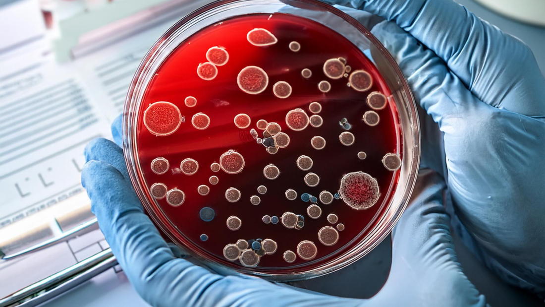
Blood culture is a critical diagnostic test used by healthcare professionals to detect infections in the bloodstream. This simple yet powerful test allows doctors to identify the presence of harmful microorganisms, such as bacteria, fungi, or parasites, that may have entered the blood, causing severe and potentially life-threatening conditions. By identifying the exact pathogen responsible for the infection, blood cultures play a pivotal role in guiding treatment decisions, ensuring patients receive the most effective care tailored to their specific condition.

What is Blood Culture?

A blood culture is a laboratory test that checks for foreign invaders like bacteria, fungi, and other pathogens in a patient’s blood. The presence of microorganisms in the bloodstream can indicate serious infections, such as sepsis, which requires immediate medical attention. Blood is normally sterile, meaning it does not contain any bacteria or pathogens. When pathogens invade the blood, they can spread rapidly to various parts of the body, leading to systemic infections.

Importance of Blood Culture in Medical Science

Blood culture serves as an essential tool in modern medicine for diagnosing infections that may be difficult to detect through other means. It provides clinicians with critical information regarding the type of pathogen causing the infection, the extent of the infection, and the best possible treatment plan. Without proper identification of the microorganism, treatment can become a trial-and-error process, which may lead to prolonged illness or even death. Blood cultures also help in tracking the effectiveness of treatment, as follow-up blood cultures can show whether the infection is responding to therapy.

How Blood Culture is Performed

The process of performing a blood culture involves drawing blood from a patient, typically from a vein in the arm. The blood is then placed in specialized bottles containing nutrients that allow microorganisms, if present, to grow. These bottles are kept under controlled laboratory conditions to encourage the growth of any pathogens. After an incubation period, laboratory technicians examine the samples to identify the type of microorganism present, which is crucial for deciding the appropriate treatment.

Hemoglobin Electrophoresis

All about Hemoglobin Electrophoresis?

About Hemoglobin Electrophoresis:-

Introduction to Hemoglobin Electrophoresis

Hemoglobin electrophoresis is a laboratory technique that separates different types of hemoglobin present in the blood. Hemoglobin is the protein responsible for carrying oxygen from the lungs to the rest of the body and returning carbon dioxide back to the lungs to be exhaled. The structure of hemoglobin can vary due to genetic mutations, resulting in different forms such as Hemoglobin A, Hemoglobin S, Hemoglobin C, Hemoglobin F, and others. These variations are crucial in diagnosing disorders such as sickle cell disease, thalassemia, and other hemoglobinopathies.

The process of hemoglobin electrophoresis allows doctors to identify these abnormal hemoglobins by analyzing the proteins’ migration patterns in an electric field. By understanding the types of hemoglobin present, healthcare providers can diagnose and monitor conditions more effectively.

Understanding Hemoglobin

Before delving into hemoglobin electrophoresis, it’s essential to understand hemoglobin itself. Hemoglobin is a globular protein found in red blood cells (RBCs) and is composed of four polypeptide chains: two alpha and two beta chains in adults, collectively referred to as Hemoglobin A (HbA). Hemoglobin’s primary role is to transport oxygen from the lungs to the tissues and return carbon dioxide from tissues to the lungs.

There are different types of hemoglobin found in the human body, including:

  • Hemoglobin A (HbA): The predominant hemoglobin in adults, consisting of two alpha and two beta chains.
  • Hemoglobin A2 (HbA2): A minor component of adult hemoglobin, made up of two alpha and two delta chains.
  • Hemoglobin F (HbF): Fetal hemoglobin, composed of two alpha and two gamma chains, is present in fetuses and newborns and gradually decreases after birth.

Types of Hemoglobin Disorders

Hemoglobinopathies are genetic conditions that result from abnormalities in the hemoglobin structure or production. The most common types of hemoglobin disorders include:

  1. Sickle Cell Disease (SCD): Caused by an abnormal form of hemoglobin called Hemoglobin S (HbS), which leads to the sickling of red blood cells. These sickled cells can cause blockages in blood vessels, leading to pain, anemia, and organ damage.
  2. Thalassemia: A group of inherited blood disorders in which the body produces an abnormal amount or form of hemoglobin. There are two main types:
    • Alpha Thalassemia: A condition in which there is a deficiency in alpha chains.
    • Beta Thalassemia: A condition in which beta chains are affected.
  3. Hemoglobin C Disease: Caused by Hemoglobin C (HbC), which leads to the production of abnormal hemoglobin and can cause mild anemia.
  4. Hemoglobin E Disease: A form of hemoglobinopathy found primarily in Southeast Asia. Hemoglobin E (HbE) leads to a mild form of anemia and, in combination with other hemoglobin mutations, can result in more severe conditions.

Principles Behind Hemoglobin Electrophoresis

The core principle of hemoglobin electrophoresis lies in the separation of hemoglobin types based on their charge and size. The different types of hemoglobin proteins have varying electrical charges, which cause them to migrate at different speeds when an electrical current is applied in a gel or cellulose medium. This allows for the identification of various hemoglobin types by comparing their migration patterns to known standards.

The Procedure of Hemoglobin Electrophoresis

  1. Sample Collection: Blood samples are collected from the patient. The red blood cells are lysed to release hemoglobin into the solution.
  2. Electrophoresis Setup: The hemoglobin sample is placed on a gel, which is placed in a buffer solution. An electrical current is applied across the gel, and the hemoglobin molecules start to migrate based on their charge and size.
  3. Migration: Different types of hemoglobin (HbA, HbS, HbC, etc.) migrate at different rates due to their different electric charges.
  4. Staining and Identification: After the migration is complete, the gel is stained to visualize the separated bands of hemoglobin. These bands are compared to a reference standard to determine the types of hemoglobin present.

Interpreting Results from Hemoglobin Electrophoresis

The results from hemoglobin electrophoresis are presented as distinct bands on the gel. Each band corresponds to a different type of hemoglobin. Based on the relative intensity and location of these bands, healthcare providers can determine which types of hemoglobin are present and in what proportions. For example:

  • Hemoglobin A (HbA): In normal adults, the predominant band will be HbA, as it makes up about 95-98% of total hemoglobin.
  • Hemoglobin F (HbF): This hemoglobin is common in newborns, but its presence in adults may indicate certain disorders like beta thalassemia or other hereditary conditions.
  • Hemoglobin S (HbS): A significant presence of HbS is indicative of sickle cell disease or sickle cell trait.
  • Hemoglobin C (HbC): The presence of HbC suggests hemoglobin C disease or trait.
  • Hemoglobin E (HbE): HbE presence points toward hemoglobin E disease, prevalent in certain regions such as Southeast Asia.

Common Uses of Hemoglobin Electrophoresis

Hemoglobin electrophoresis is essential for diagnosing several conditions related to abnormal hemoglobin, including:

  • Sickle Cell Disease: Identifying HbS can confirm sickle cell disease or sickle cell trait.
  • Thalassemia: The test is useful in detecting abnormal proportions of HbA2 and HbF, which are indicative of alpha or beta thalassemia.
  • Hemoglobin C and E Disorders: The test helps in identifying individuals who may have hemoglobin C or E traits or related diseases.
  • Prenatal Screening: In some cases, hemoglobin electrophoresis is used in prenatal screening to assess the risk of hemoglobinopathies in a fetus.

Why is Hemoglobin Electrophoresis Important?

The test plays a critical role in the diagnosis and management of hemoglobinopathies. Many of these conditions are inherited and can be passed on to children, making early detection important for family planning and treatment decisions. For individuals with sickle cell disease or thalassemia, managing these conditions often involves lifelong care. Early diagnosis through hemoglobin electrophoresis enables patients to receive timely treatment, which can significantly improve their quality of life.

Additionally, hemoglobin electrophoresis is often part of newborn screening programs, particularly in regions where hemoglobinopathies are common. Early detection in newborns allows for early intervention, which can prevent serious complications later in life.


This introduction can be greatly expanded by diving deeper into each of the following:

  1. Historical Overview of Hemoglobin Electrophoresis
    • Early discoveries and breakthroughs in hemoglobin analysis.
    • Development of electrophoresis as a diagnostic tool.
  2. Technical Aspects of Electrophoresis
    • The chemistry behind hemoglobin separation.
    • Equipment used and advancements in technology.
    • Detailed exploration of the buffer systems and gels used.
  3. Genetic Aspects of Hemoglobin Disorders
    • How mutations lead to different hemoglobin types.
    • Inheritance patterns of hemoglobinopathies.
    • Global distribution of hemoglobin disorders (prevalence in specific regions).
  4. Clinical Applications
    • Case studies showcasing real-world applications of hemoglobin electrophoresis.
    • How treatment plans change based on electrophoresis results.
    • Emerging research on hemoglobin variants and their health implications.
  5. Emerging Technologies
    • Advances in electrophoresis methods.
    • Automated systems and their role in clinical laboratories.
    • The future of hemoglobin diagnostics with molecular techniques.

You can check this product :- Click the below product link.

MiniCap Flex-Piercing