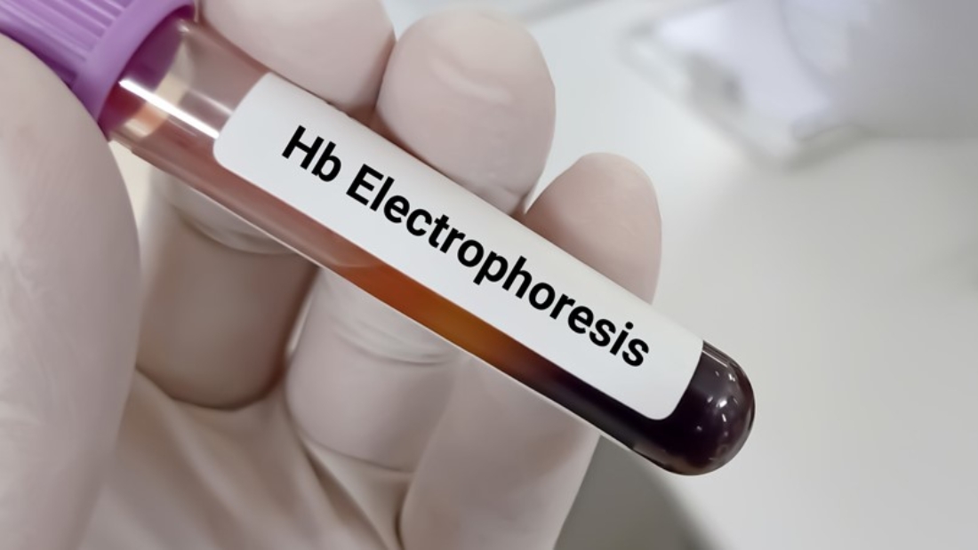
About Hemoglobin Electrophoresis:-
Introduction to Hemoglobin Electrophoresis
Hemoglobin electrophoresis is a laboratory technique that separates different types of hemoglobin present in the blood. Hemoglobin is the protein responsible for carrying oxygen from the lungs to the rest of the body and returning carbon dioxide back to the lungs to be exhaled. The structure of hemoglobin can vary due to genetic mutations, resulting in different forms such as Hemoglobin A, Hemoglobin S, Hemoglobin C, Hemoglobin F, and others. These variations are crucial in diagnosing disorders such as sickle cell disease, thalassemia, and other hemoglobinopathies.
The process of hemoglobin electrophoresis allows doctors to identify these abnormal hemoglobins by analyzing the proteins’ migration patterns in an electric field. By understanding the types of hemoglobin present, healthcare providers can diagnose and monitor conditions more effectively.
Understanding Hemoglobin
Before delving into hemoglobin electrophoresis, it’s essential to understand hemoglobin itself. Hemoglobin is a globular protein found in red blood cells (RBCs) and is composed of four polypeptide chains: two alpha and two beta chains in adults, collectively referred to as Hemoglobin A (HbA). Hemoglobin’s primary role is to transport oxygen from the lungs to the tissues and return carbon dioxide from tissues to the lungs.
There are different types of hemoglobin found in the human body, including:
- Hemoglobin A (HbA): The predominant hemoglobin in adults, consisting of two alpha and two beta chains.
- Hemoglobin A2 (HbA2): A minor component of adult hemoglobin, made up of two alpha and two delta chains.
- Hemoglobin F (HbF): Fetal hemoglobin, composed of two alpha and two gamma chains, is present in fetuses and newborns and gradually decreases after birth.
Types of Hemoglobin Disorders
Hemoglobinopathies are genetic conditions that result from abnormalities in the hemoglobin structure or production. The most common types of hemoglobin disorders include:
- Sickle Cell Disease (SCD): Caused by an abnormal form of hemoglobin called Hemoglobin S (HbS), which leads to the sickling of red blood cells. These sickled cells can cause blockages in blood vessels, leading to pain, anemia, and organ damage.
- Thalassemia: A group of inherited blood disorders in which the body produces an abnormal amount or form of hemoglobin. There are two main types:
- Alpha Thalassemia: A condition in which there is a deficiency in alpha chains.
- Beta Thalassemia: A condition in which beta chains are affected.
- Hemoglobin C Disease: Caused by Hemoglobin C (HbC), which leads to the production of abnormal hemoglobin and can cause mild anemia.
- Hemoglobin E Disease: A form of hemoglobinopathy found primarily in Southeast Asia. Hemoglobin E (HbE) leads to a mild form of anemia and, in combination with other hemoglobin mutations, can result in more severe conditions.
Principles Behind Hemoglobin Electrophoresis
The core principle of hemoglobin electrophoresis lies in the separation of hemoglobin types based on their charge and size. The different types of hemoglobin proteins have varying electrical charges, which cause them to migrate at different speeds when an electrical current is applied in a gel or cellulose medium. This allows for the identification of various hemoglobin types by comparing their migration patterns to known standards.
The Procedure of Hemoglobin Electrophoresis
- Sample Collection: Blood samples are collected from the patient. The red blood cells are lysed to release hemoglobin into the solution.
- Electrophoresis Setup: The hemoglobin sample is placed on a gel, which is placed in a buffer solution. An electrical current is applied across the gel, and the hemoglobin molecules start to migrate based on their charge and size.
- Migration: Different types of hemoglobin (HbA, HbS, HbC, etc.) migrate at different rates due to their different electric charges.
- Staining and Identification: After the migration is complete, the gel is stained to visualize the separated bands of hemoglobin. These bands are compared to a reference standard to determine the types of hemoglobin present.
Interpreting Results from Hemoglobin Electrophoresis
The results from hemoglobin electrophoresis are presented as distinct bands on the gel. Each band corresponds to a different type of hemoglobin. Based on the relative intensity and location of these bands, healthcare providers can determine which types of hemoglobin are present and in what proportions. For example:
- Hemoglobin A (HbA): In normal adults, the predominant band will be HbA, as it makes up about 95-98% of total hemoglobin.
- Hemoglobin F (HbF): This hemoglobin is common in newborns, but its presence in adults may indicate certain disorders like beta thalassemia or other hereditary conditions.
- Hemoglobin S (HbS): A significant presence of HbS is indicative of sickle cell disease or sickle cell trait.
- Hemoglobin C (HbC): The presence of HbC suggests hemoglobin C disease or trait.
- Hemoglobin E (HbE): HbE presence points toward hemoglobin E disease, prevalent in certain regions such as Southeast Asia.
Common Uses of Hemoglobin Electrophoresis
Hemoglobin electrophoresis is essential for diagnosing several conditions related to abnormal hemoglobin, including:
- Sickle Cell Disease: Identifying HbS can confirm sickle cell disease or sickle cell trait.
- Thalassemia: The test is useful in detecting abnormal proportions of HbA2 and HbF, which are indicative of alpha or beta thalassemia.
- Hemoglobin C and E Disorders: The test helps in identifying individuals who may have hemoglobin C or E traits or related diseases.
- Prenatal Screening: In some cases, hemoglobin electrophoresis is used in prenatal screening to assess the risk of hemoglobinopathies in a fetus.
Why is Hemoglobin Electrophoresis Important?
The test plays a critical role in the diagnosis and management of hemoglobinopathies. Many of these conditions are inherited and can be passed on to children, making early detection important for family planning and treatment decisions. For individuals with sickle cell disease or thalassemia, managing these conditions often involves lifelong care. Early diagnosis through hemoglobin electrophoresis enables patients to receive timely treatment, which can significantly improve their quality of life.
Additionally, hemoglobin electrophoresis is often part of newborn screening programs, particularly in regions where hemoglobinopathies are common. Early detection in newborns allows for early intervention, which can prevent serious complications later in life.
This introduction can be greatly expanded by diving deeper into each of the following:
- Historical Overview of Hemoglobin Electrophoresis
- Early discoveries and breakthroughs in hemoglobin analysis.
- Development of electrophoresis as a diagnostic tool.
- Technical Aspects of Electrophoresis
- The chemistry behind hemoglobin separation.
- Equipment used and advancements in technology.
- Detailed exploration of the buffer systems and gels used.
- Genetic Aspects of Hemoglobin Disorders
- How mutations lead to different hemoglobin types.
- Inheritance patterns of hemoglobinopathies.
- Global distribution of hemoglobin disorders (prevalence in specific regions).
- Clinical Applications
- Case studies showcasing real-world applications of hemoglobin electrophoresis.
- How treatment plans change based on electrophoresis results.
- Emerging research on hemoglobin variants and their health implications.
- Emerging Technologies
- Advances in electrophoresis methods.
- Automated systems and their role in clinical laboratories.
- The future of hemoglobin diagnostics with molecular techniques.
You can check this product :- Click the below product link.


